The Cow Eye Dissection Lab Worksheet Answers
Biology A Cow Eye Dissection Purpose To Gain An
Cow Eye Dissection
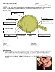
Cow Eye Dissection Lab Cow Eye Dissection Lab Name Date
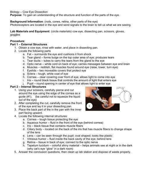
Terms in this set 17 aqueous humor.
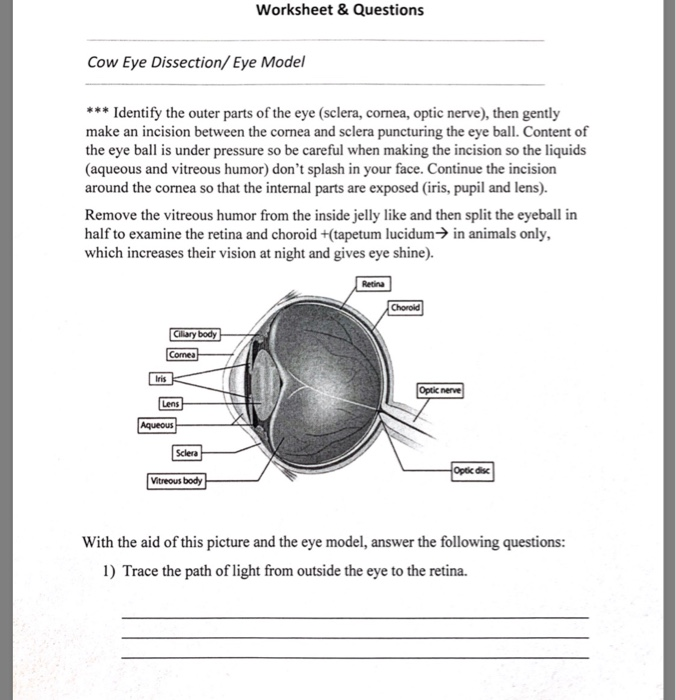
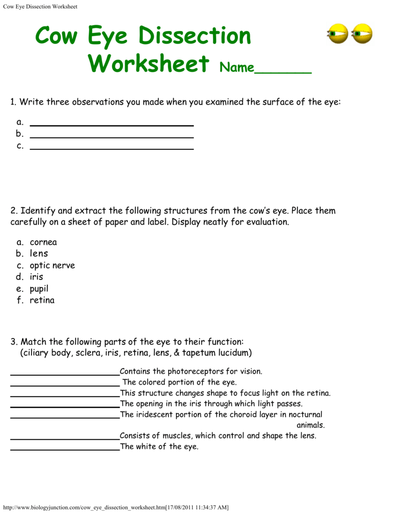
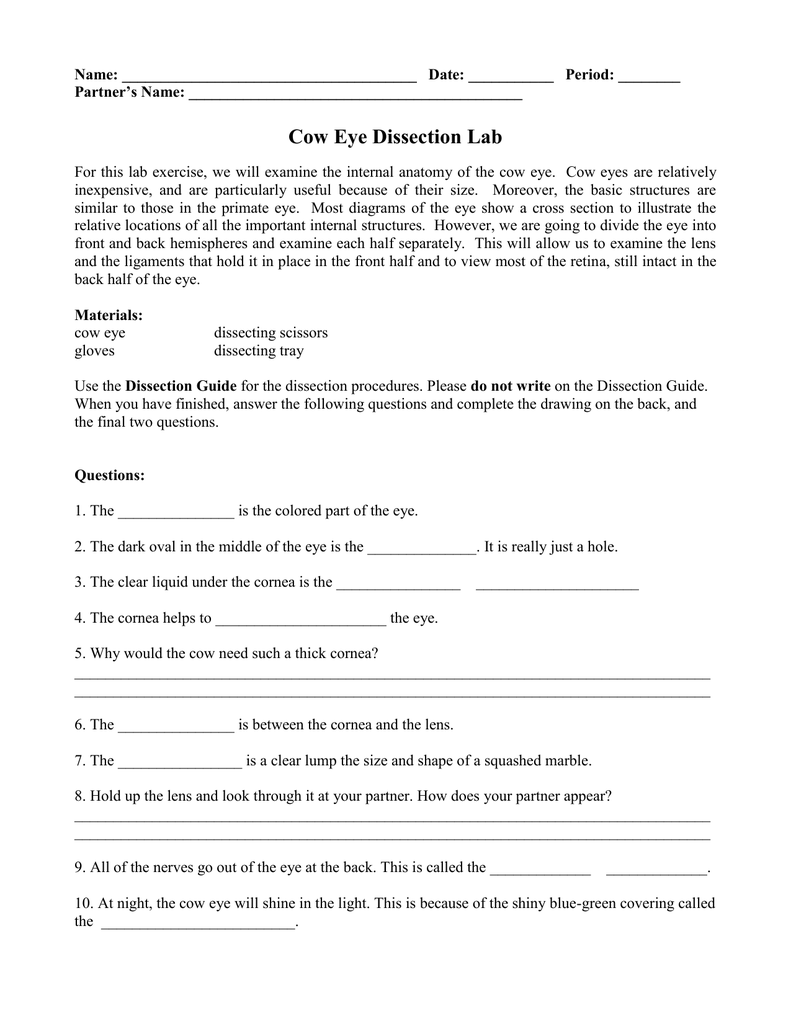
The cow eye dissection lab worksheet answers. Answer the worksheet questions on the cow eye dissection. Clear fluid filling the area between the lens and cornea composed mostly of water. Examine the outside of the eye. The cow eye dissection lab what are the structures of the mammalian eye and how do they function.
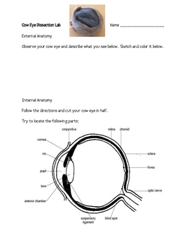
Sends messages from the eye to the brain. Name the three layers you sliced through when you cut across the top. Carbon which so many of us take for granted is actually one. Identify the following structures.
Cows eye dissection page 6 now take a look at the rest of the eye. The structures have certain functions and together they form images that are interpreted by the brain. Cow eye dissection. This was a question that we felt deserved an in depth answer.
Tell three observations you made when you examined the surface of the eye. The mammalian eye consists of many specialized cells and tissues that make up several different structures. Helps maintain the shape of your eyeball. On the inside of the back half of the eyeball you can see some blood.
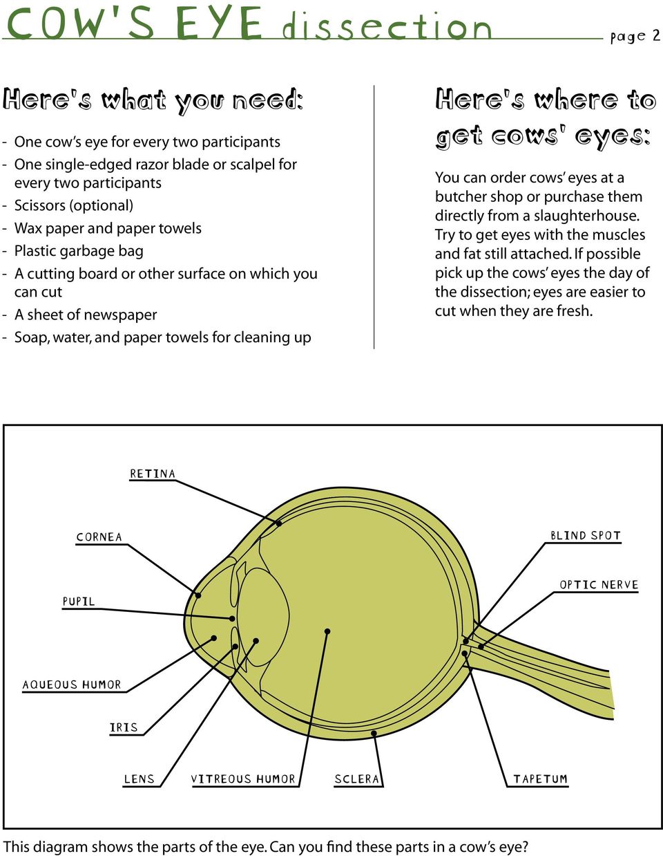
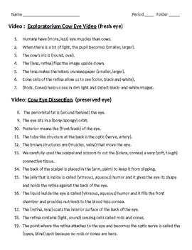
Hole in the iris that allows light into the inner eye. The cow eye is a fantastic specimen for students of all ages to dissect. Click here for eye dissection questions. The lab guide for students outlines the procedure for the dissection and you can view the eye gallery to see photographs of the dissection.
The cow eye dissection lab what are the structures of the mammalian eye and how do they function. The structures have certain functions and together they form images that are interpreted by the brain. You should be able to find the sclera or the whites of the eye. The mammalian eye consists of many specialized cells and tissues that make up several different structures.
2. Inside and outside of the eye and helps maintain the shape of the eye. Cornea tear gland optic nerve iris pupil retina 3. A layer of cells in the back of the eye that picks up vibrations of visible light.
If the vitreous humor is still in the eyeball empty it out. Cow eye dissection worksheet 1. Contains detailed instructions images and an image for labeling the parts of the eye such as the retina tapetum and optic nerve. The structures are clear dissection easy to accomplish and usually kids enjoy the lab.
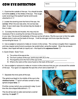
Procedure cornea optic nerve fat muscle sclera cornea procedure continued on the next page. This tough outer. Also it is the attachment point of the muscles that allow the eye to move. Student lab guide for dissecting a cow or a sheep eye.
Colored ring of muscle that changes the size of the pupil.

Cow Eye Dissection Worksheet Cow Eye Dissection Directions
Cow Eye Dissection Lab For Elementary

Cow Eye Dissection Worksheet Scaffolded
Cow Eye Dissection Worksheet

Anatomy Of Eye Worksheets Worksheets Cow Eye Dissection
Cow Eye Dissection
Cow Eye Dissection Worksheets Teaching Resources Tpt
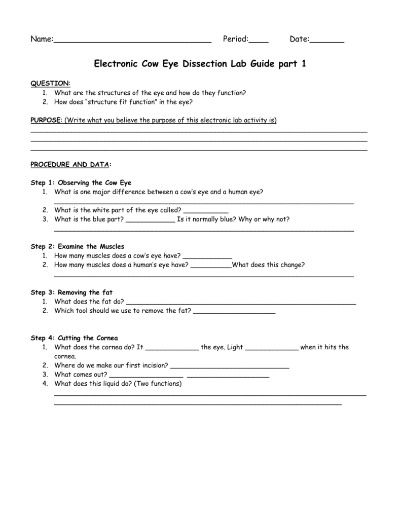
Electronic Cow Eye Dissection Lab Guide Part 1

Cow Eye Dissection Worksheet Cow Eyes Biology Eye Anatomy

Cow Eye Dissection Or What Else Do You Do On A Saturday

Cow Eye Dissection

Cow Eye Dissection Or What Else Do You Do On A Saturday
Cow S Eye Dissection Dissecting A Cow S Eye Step By Step
Cow Eye Dissection Worksheets Teaching Resources Tpt
Cow Eye Dissection Lab

Cow S Eye Dissection Eye Diagram

Lab Virtual Cow Eye Dissection Mh Docx Lab Virtual Eye

Cow S Eye Dissection Eye Diagram Cow Eyes Human Anatomy
